
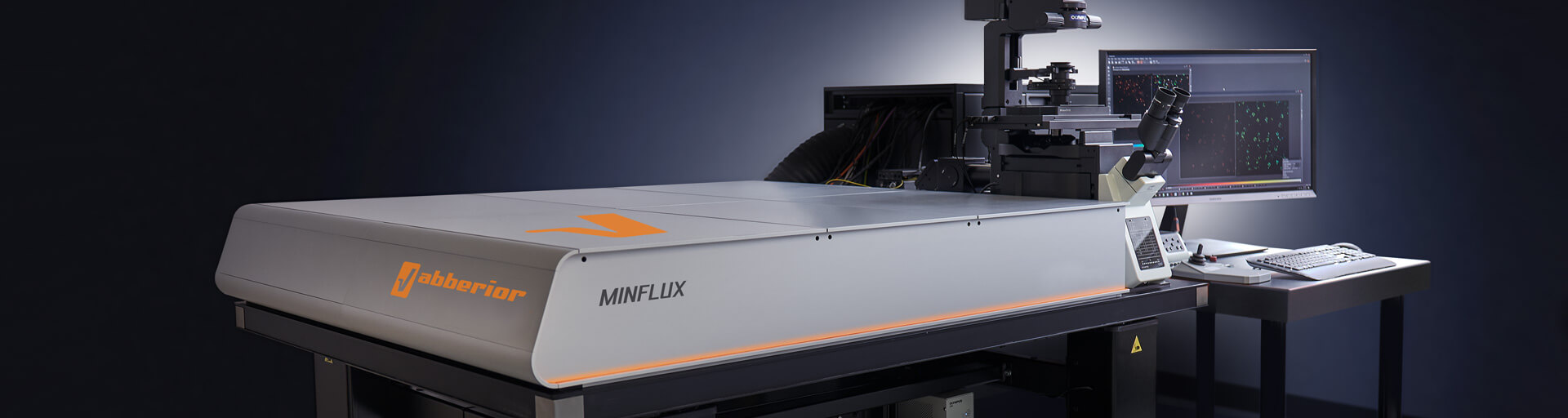
Abberior MINFLUX
MINimal flourescence photon FLUXes nanoscopy
Traditional fluorescence microscopy is limited by the diffraction limit of light, which restricts resolution to about 200-300 nanometers. MINFLUX breaks this limit and allows imaging at much higher resolutions (down to a few nanometers).
© 2024 abberior
MINFLUX
- Tracks labeled molecules at high frequencies up to 10 kHz, resolving molecular motion every 100 μs
- Achieves localization precisions of 1-3 nm (3D) in biological samples on large fields of view (10 x 15 μm)
- Compatible with both live and fixed fluorescently labeled specimens. Multi-color imaging is possible.
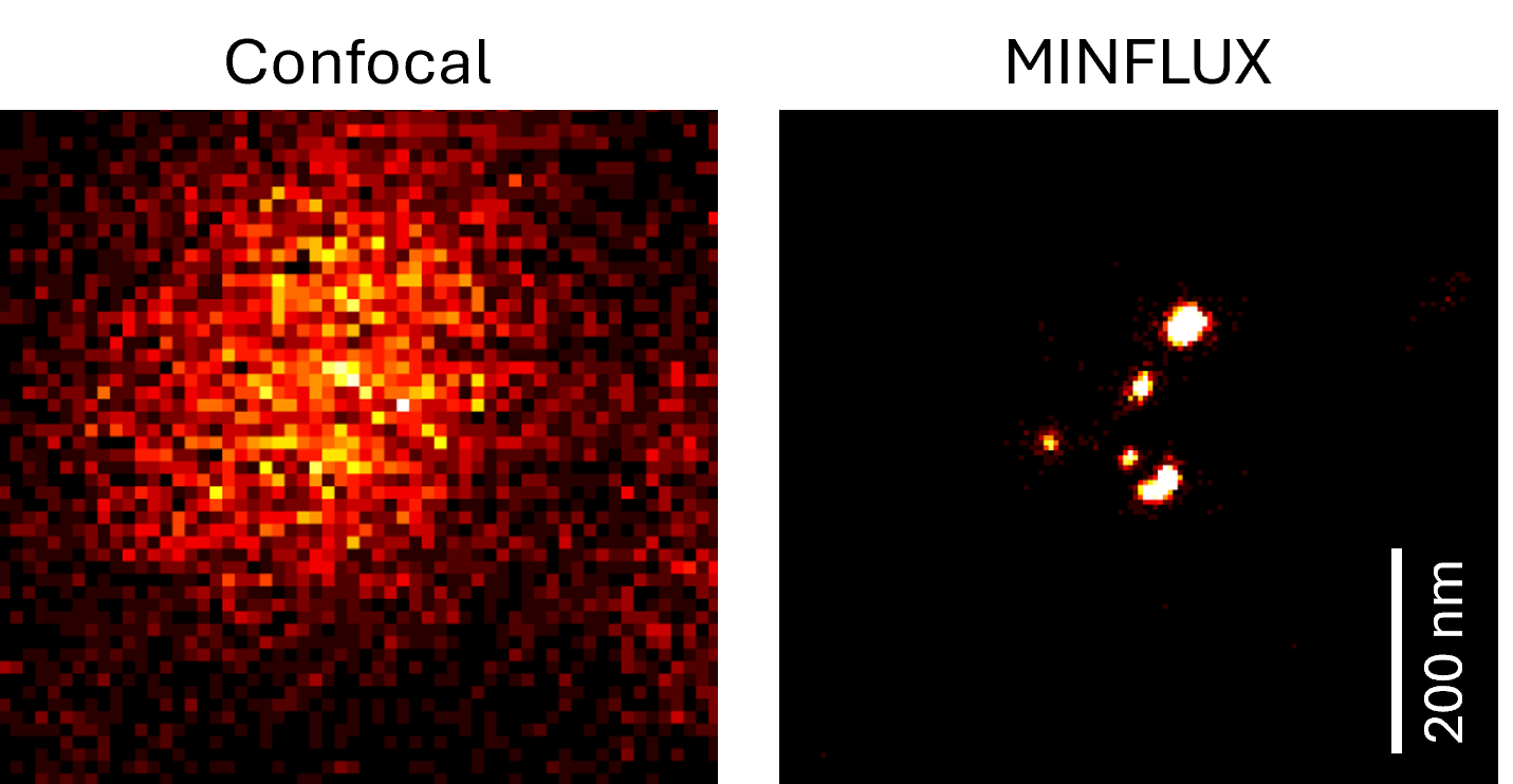
Comparison between confocal and MINFLUX resolution. Individual RNA probes hybridized along an RNA strand. © 2024 DaMBIC.
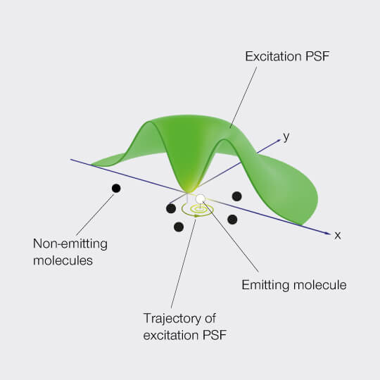
MINFLUX principle
MINFLUX works by the principle of minimal photon fluxes where fluorophore positions can be determined with a minimal number of photons and consequently within a fast and accurate spatio-temporal regime. The single emitting molecules are localized with an excitation pattern featuring a spatially well-controlled intensity zero, e.g. a donut as seen below. The intensity zero of this excitation beam is then scanned onto the sample and used to probe the fluorophore positions.
© 2024 abberior
System details
Resolution down to 1-3 nm
The system contains two pulsed excitation laser sources @ 561 nm and @ 640 nm allowing for imaging of fluorophores in the excitation range ~550 - 680 nm.
The MINFLUX is built onto an IX83 Olympus microscope with a multicolor CoolLED illumination source.
Location: V12-605b-0